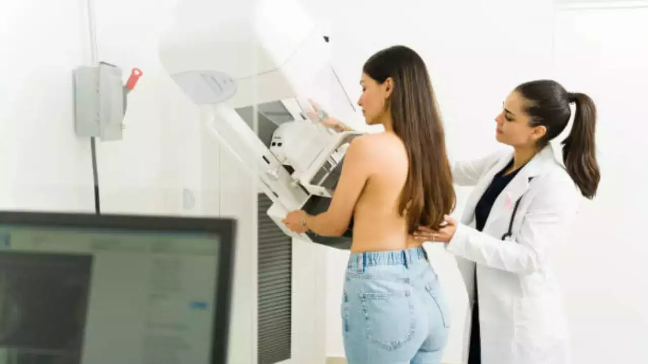
Imaging by means of radiation medicine techniques is the first step in clinical management and diagnostic radiology
Cancer management has never been easy. Not only does it require a reliable diagnosis to identify the primary tumour and assess its dissemination to surrounding tissues, but a need to check the other organs and structures throughout the body is also there. Imaging by means of radiation medicine techniques is the first step in clinical management and diagnostic radiology and nuclear medicine studies play important roles in screening, staging, and monitoring of treatment.
However, until a few decades ago, medical imaging was dominated by projection-view X-ray radiography aimed at detecting changes in tissue density. However, with the continuous advancement in science and improvements in computer technology, many kinds of digital techniques have been introduced into medical radiation imaging for foolproof diagnosis.
“Beginning from the invention of X-rays and moving into positron emission tomography (PET) scans and the addition of intelligence in the type of artificial intelligence (AI), imaging has had a vital part to play in the changing face of cancer care,” Dr. Siddharth Parekh, Consultant Radiology, HCG Cancer Centre, told Times Now.
How did the journey begin from X-rays
Radiography was the first imaging technique applied in oncology and it yielded crucial data regarding the basic internal structure of the body. However, according to Dr. Parekh, X-rays have a few drawbacks. “For instance, X-ray images cannot produce anatomical structure details in an adequate manner or distinguish between the types of tissue,” he said.
This occasionally resulted in the inability to differentiate between malignant and non-cancerous tissues, emphasizing the importance of better imaging methods.
The introduction of Magnetic Resonance Imaging or MRI
MRI has thereafter been one of the most significant developments in oncology imaging as it provides field images with notably great detail, enabling physicians to distinguish between normal and malignant tissue.
“This allows specific prevalence and treatment planning, which makes MRI critical in cancer diagnosis,” said Dr. Parekh.
The emergence of Positron Emission Tomography or PET scans
PET has introduced functional imaging in oncology care, making it possible to assess the metabolic activity of tumours.
“PET scans are able to identify regions of menacing cell activity, recognize the scope of the disease, and judge the efficacy of the therapy. In many situations, PET scans can also identify the place of origin of cancer and therefore form an important part of cancer staging and diagnosis,” Dr. Parekh added.
What is the future of oncology imaging?
According to Dr. Parekh, there are several categories of molecular imaging, like fluorescence imaging and targeted radiotracers, which extend the impact on oncology imaging. These methods not only allow positive molecules to be visualized but also yield useful information about the biological activity of a tumour.
Experts believe that by identifying the tumours at the molecular level, it becomes easier for a physician to diagnose and treat the patients, hence enhancing patient care.
How does AI integrate into oncology imaging?
There are several uses of artificial intelligence in oncology imaging with huge potential. Dr. Parekh explains that AI can be useful in processing large volumes of data acquired from imaging that may contain subtle changes that can only be spotted using AI.
“This technology may offer a way to increase early cancer detection, help with diagnosis, and evaluate the leading factors with regard to a patient’s response to treatment,” he said.
Get Latest News Live on Times Now along with Breaking News and Top Headlines from Health and around the world.